Facilities & Techniques
Research Facilities
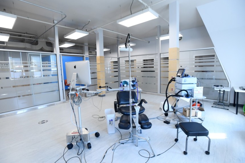
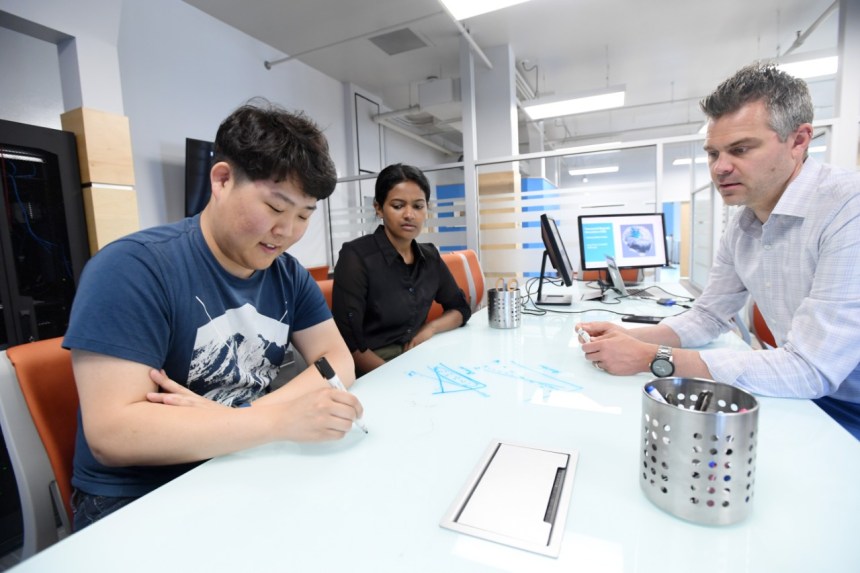
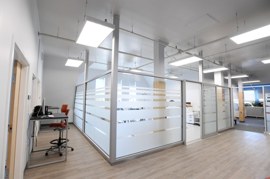
The Laboratory for Brain Recovery and Function comprises ~2400 ft2, including 3 individual lab rooms, a large (~500 ft2) collaborative student space and a common experimental room (~500 ft2).
In addition to state-of-the-art equipment (see research techniques below), the CFI-funded laboratory has capacity for analysis of large-scale data using an in-house server featuring 24 processing cores, 384 GB RAM and 26 TB storage, which is complemented by a file-exchange server with high capacity (8TB) storage.
Here’s a video walk-through of the Laboratory for Brain Research and Function
Research Techniques
Work in the lab encompasses a number of experimental techniques including electrophysiological and neuroimaging methods, in addition to behaviour-based outcome measures to investigate questions related to the function of the human nervous system in health and disease. In addition to those detailed below, students in the lab have access to a number of other techniques through our collaborators, including functional near infrared spectroscopy (fNIRS) and portable EEG. Through the School of Physiotherapy, students are also able to access various cycle ergometers and gas exchange units which are used in our studies examining the impact of aerobic exercise on brain function.
Studies undertaken by the lab may utilize one or more of the following techniques:
Electroencephalography (EEG)
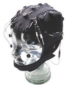 EEG is a technique that allows for the electrical activity generated by the brain to be measured. Studying the electrical activity that results from doing certain tasks can tell us about the parts of the brain that are involved in the task, or even the network of brain regions that are working together.
The Laboratory for Brain Recovery and Function is equipped with an EEG system that can measure up to 128 electrodes at any one time. The number of electrodes that are used depends on how much brain activity we want to measure (the whole brain or just parts of it), and whether we want to try to determine where the brain activity is coming from.
This latter type of analysis requires more advanced analysis techniques (called source localization) and lots of electrodes. The laboratory also has equipment that allows for digitization of the head so we can look at brain activity relative to a person’s MRI, which is a ‘picture’ of their brain.
Magnetic Resonance Imaging (MRI)
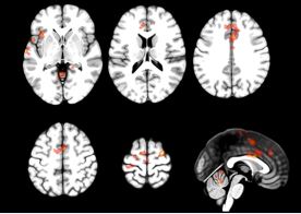 MRI is a technique used to obtain high-quality images of the body, including the brain. MRI machines use high strength magnetic fields and radio waves to obtain these images.
One kind of MRI scan that can be performed is called functional MRI (fMRI). fMRI measures blood flow in the brain in order to indirectly assess how much, and to what extent, areas of the brain are activated. fMRI is a useful tool for examining what brain areas are active when people are performing certain tasks. Other types of MRI scans can provide different kinds of information about the brain, such as looking at the connectivity (white matter) in the brain.
MRI scans performed for studies done in the lab are typically done at either 1.5T (IWK Health Centre) or 3T (Halifax Infirmary through the Biomedical Translational Imaging Centre).
Surface and/or Intramuscular Electromyography (EMG)
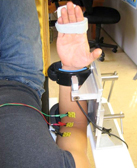 EMG is a technique that allows for the measurement of the electrical activity of muscle. EMG studies can tell us a lot about the health of the neuromuscular system, including the nerve, muscle and the nerve/muscle connection.
The Laboratory for Brain Recovery and Function is equipped with two data acquisition systems that can accommodate up to 20 channels of EMG. The lab also has equipment that allows for the stimulation of peripheral nerves, and measurement of force.
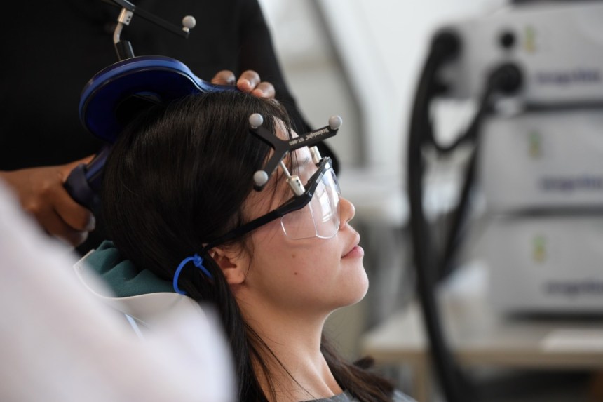
Neuro-Navigated Transcranial Magnetic Stimulation (TMS)
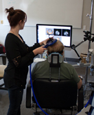 Transcranial magnetic stimulation (TMS) is a technique that permits the safe and non-invasive stimulation of the brain in intact, awake human subjects. This capacity to stimulate the human brain in a non-invasive and painless manner permits an array of opportunities that span the fields of basic and clinical neurosciences.
Three general applications of TMS have been identified, including:
1) assessment of physiological structures;
2) disruption of brain regions (so-called ‘virtual lesion’ studies); and
3) altering cortical excitability (e.g., facilitation or inhibition).
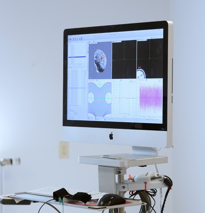
The Laboratory for Brain Recovery and Function is equipped with three TMS systems that allow for the full range of TMS applications to be performed. In addition to the TMS systems, the lab has a neuronavigation unit that pairs with the TMS to focally stimulate brain areas with anatomic specificity. The neuronavigation unit determines the position and orientation of the TMS coil relative to digitized anatomical landmarks co-registered with anatomical or functional MRI, allowing targeted stimulation with high spatial resolution.
Magnetoencephalography (MEG)
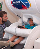 MEG is a technique that measures small magnetic fields generated by the electrical activity in the brain. These magnetic fields are representative of brain activity and can tell us about which parts of the brain are working during performance of a task and how the parts of the brain are working together.
MEG is a very useful technique because there is little set-up required and it is fairly quick to perform. Because of this it is a very ‘patient-friendly’ technique. MEG scans for studies in the lab are performed at the Laboratory for Clinical Magnetoencephalography at the IWK Health Centre.
